Basic Information
Certification
CE, FDA, ISO13485
Monitor Size
27 Inches/32 Inches
Display Resolution
1920*1080P
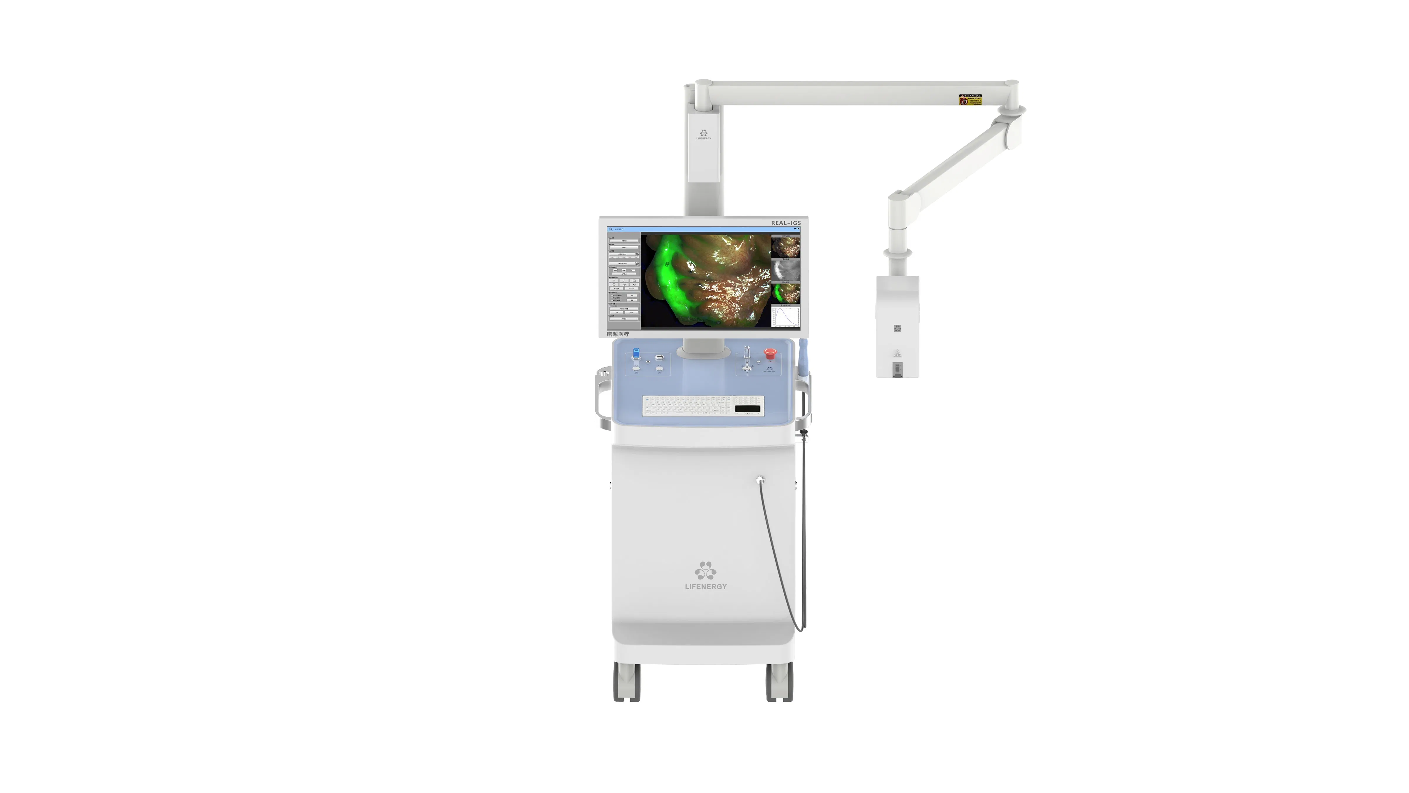
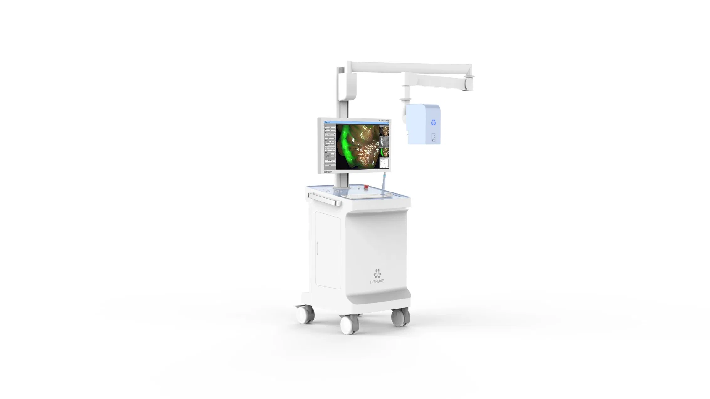
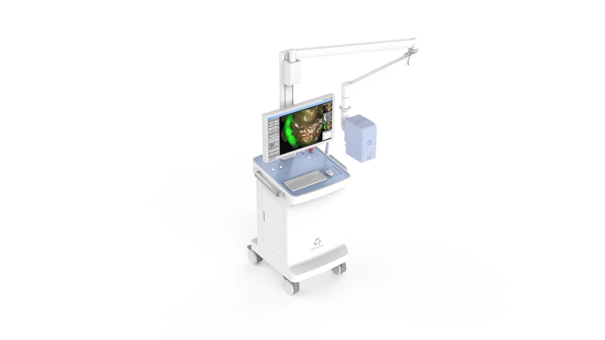
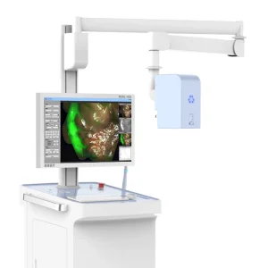
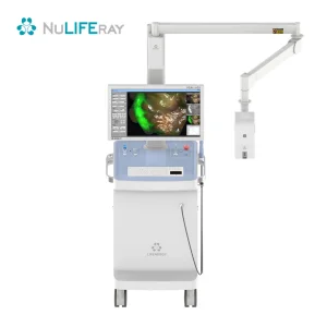
Surgical Fluorescence Imaging System
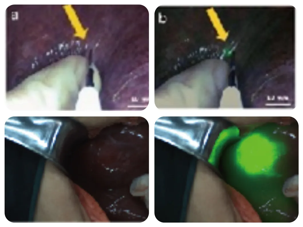
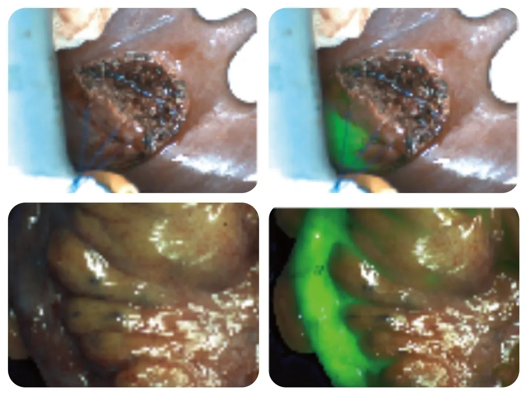
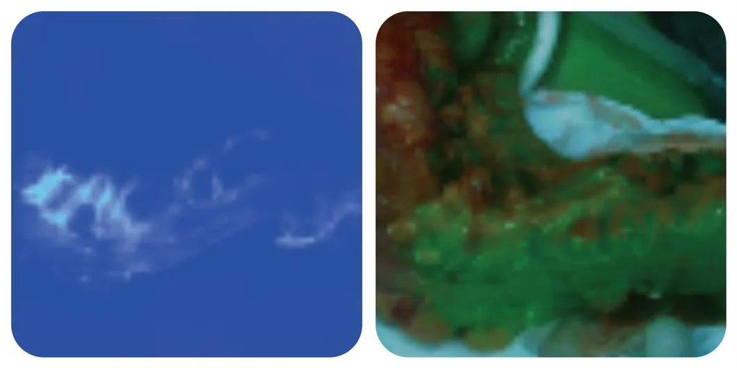
The Surgical Fluorescence Imaging System is a surgical guidance system that uses a drug-device combination approach. It employs indocyanine green (ICG) as a fluorescence probe, relying on "ultra-high sensitivity" and combining the optical properties of ICG in submillimeter-size tumors to provide surgeons with high-definition visible light, fluorescence imaging, and quantitative data for diagnostic information during tumor surgery. It is suitable for real-time observation of tissues (such as tumor tissue, margin tissue), blood supply (free skin flap), lymph nodes (sentinel lymph nodes, regional lymph nodes), and anatomical structures (liver segments, gallbladder, lung segments).
📊 Product Parameters
| Project |
Content |
| Number of Camera Chips | 2CMOS |
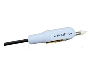
| Convenient Handheld Probe | Handheld Spectral Quantitative Analysis Probe |
| Special Fluorescence Development | AI Assisted Boundary Sharpening |
| Image Mode | 7 Types: White Light/Fluorescence/Fusion/Multimode/ColorGrading/Quantification/Spectroscopy |
| Laser Wavelength | 785nm |
| Is There a Workstation Available | Yes |
| Workstation Function | Fluorescence Intensity & Spectral Quantitative Analysis |
| Fluorescence Detection Limit | 10-12 M/L |
| Lens Zoom Factor | 4 Times |
| Camera Working Distance | Recommended 10-25cm |
| Focus Mode | Electric Focus |
| White Balance Method | Manual White Balance |
| Laser Grade | 3R |
| Is It an Integrated Model | Yes |
| Resolution of Recording and Broadcasting | High Definition |
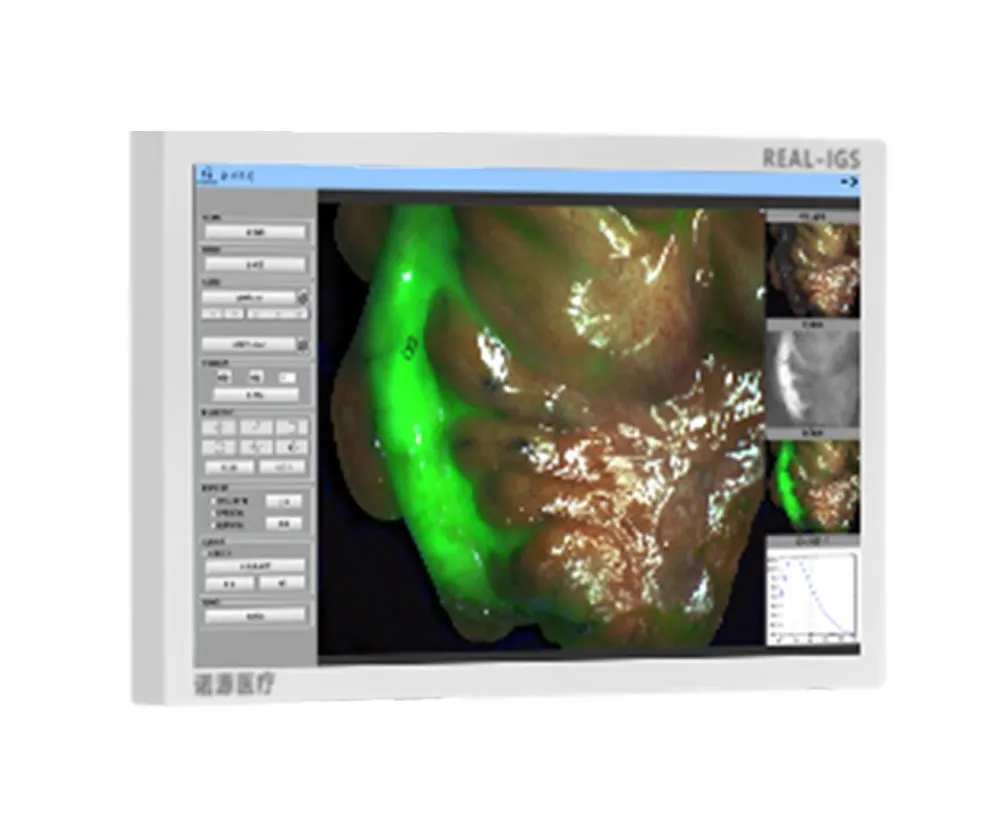
Software screenshot on the monitor
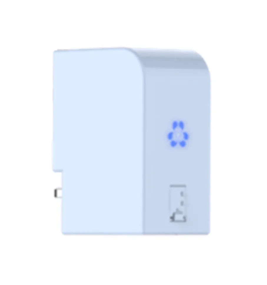
Surface light source
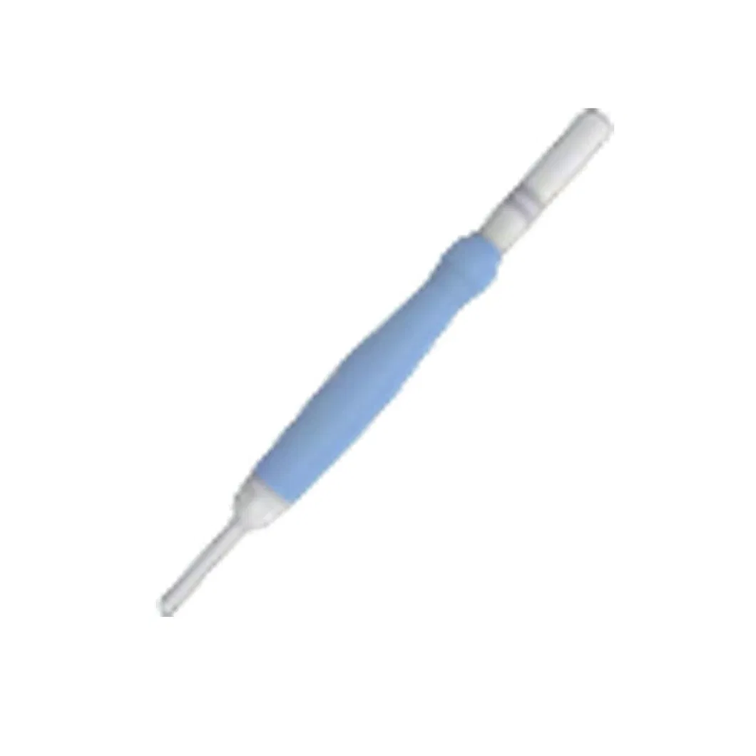
Handheld laser probe
Technical & Product Advantages
-
1
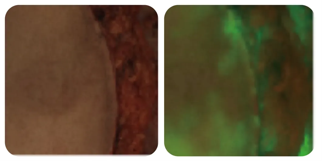
Ultra-low fluorescence detection limit: Real-time diagnosis of millimeter-sized tumors during surgery, decreasing detection limits to sub-millimeter levels.
-
2
Dual Quantitative Analysis: ROI Value and Independent Spectral Digitization provide objective numerical judgment for imaging diagnostic information.
-
3
Pseudo-color grading analysis system: Objectively presents the concentration gradient of the tracer in target tissue.
-
4
Multi-Mode Imaging: Support for 7 types including AI Assisted Boundary Sharpening and Real-time fusion.
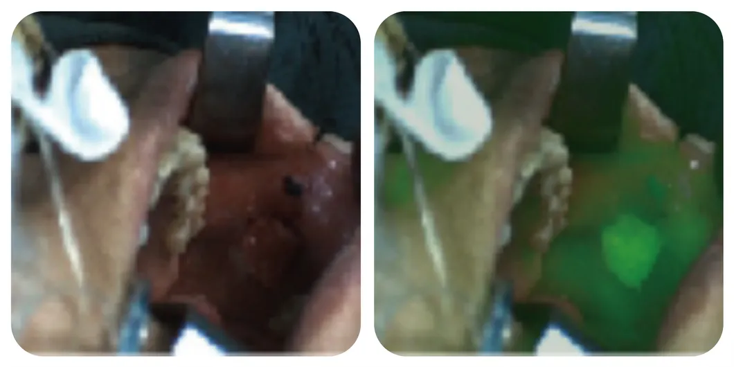
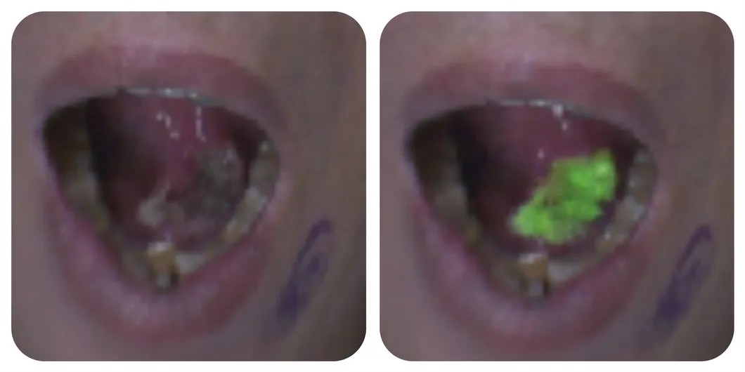
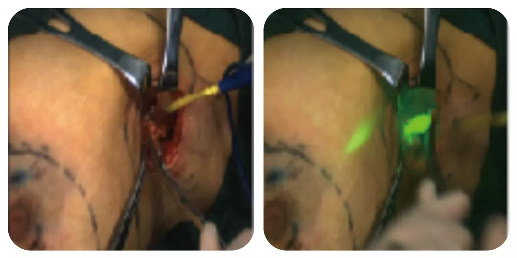
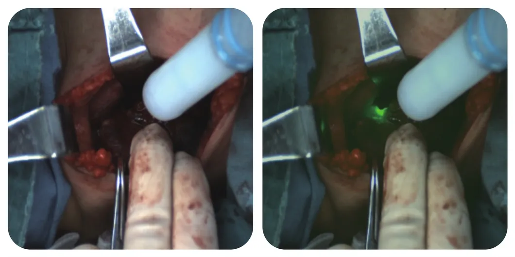
🦷 Precision Oral and Maxillofacial Surgery
Used for tumor localization, margin assessment, blood supply to transplanted flaps, and sentinel lymph node localization.
Tumor localization
Margin assessment
Blood supply assessment
Research hotspot
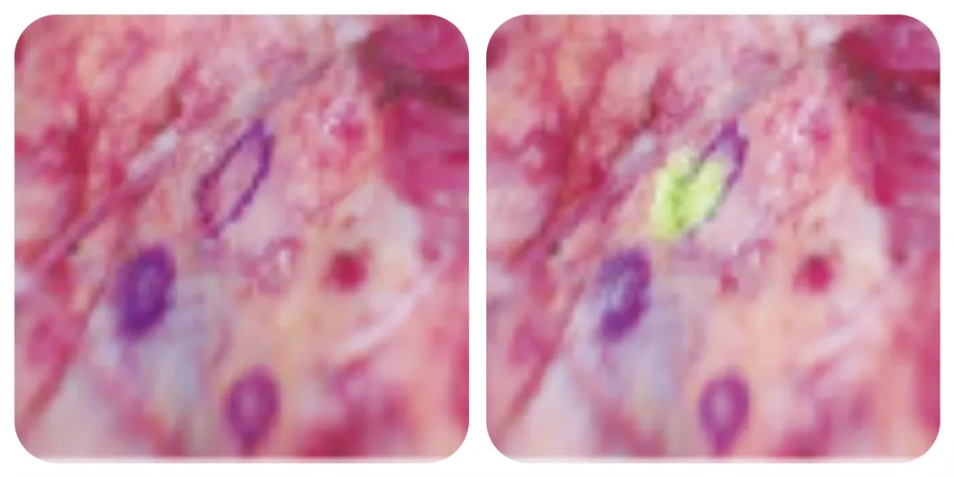
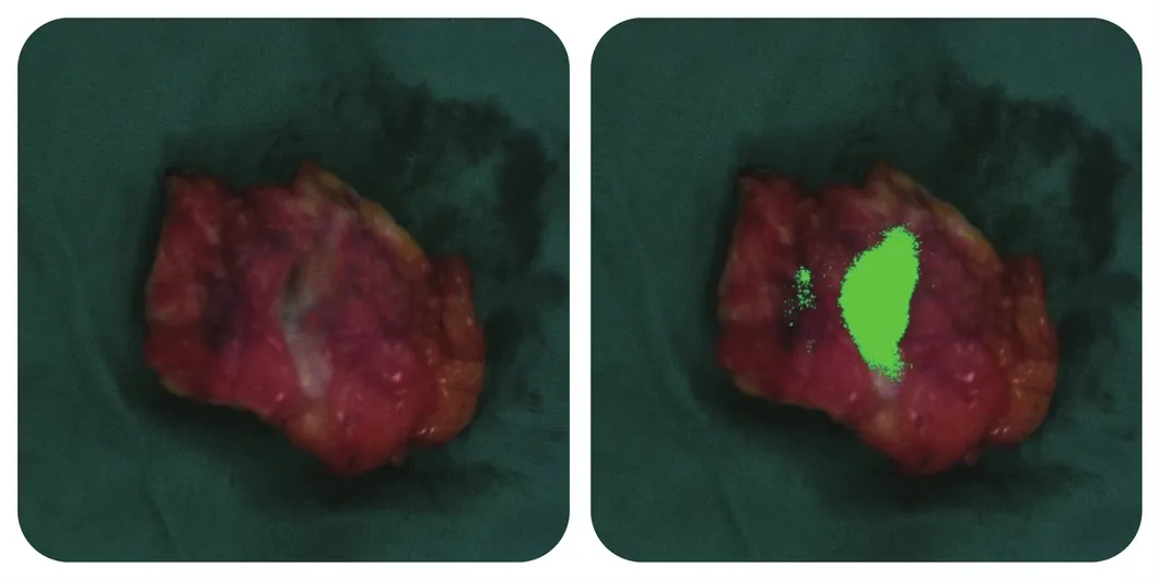
🎗️ Precision Thyroid and Breast Surgery
Identification of parathyroid glands, lymphatic mapping of sentinel nodes, and real-time tumor identification during breast-conserving surgery.
Lymphatic mapping
Tumor identification
Gland identification
Margin assessment
Functional evaluation
Blood supply evaluation
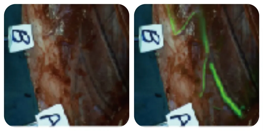
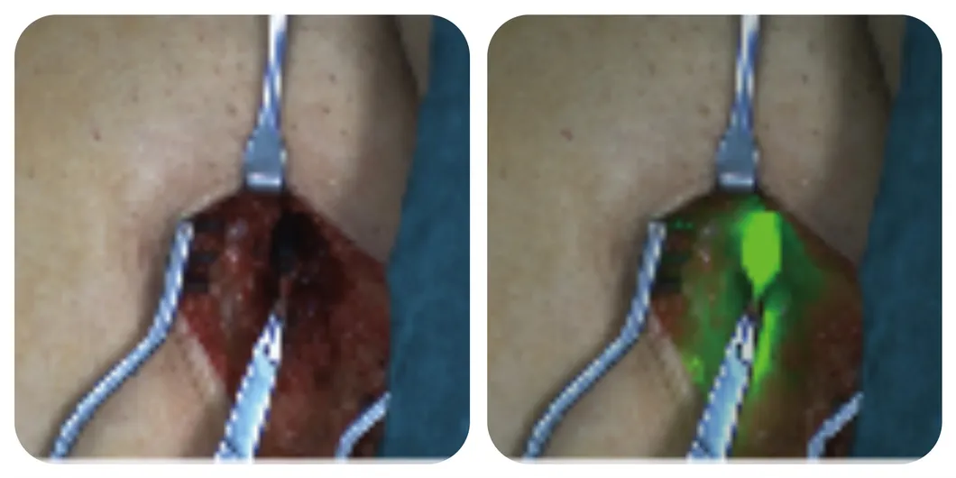
🩹 Precision Dermatology, Burns and Plastic Surgery
Lower limb lymphatic mapping
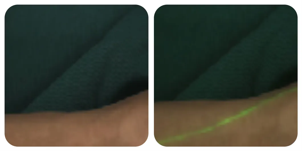
Upper limb lymphatic mapping
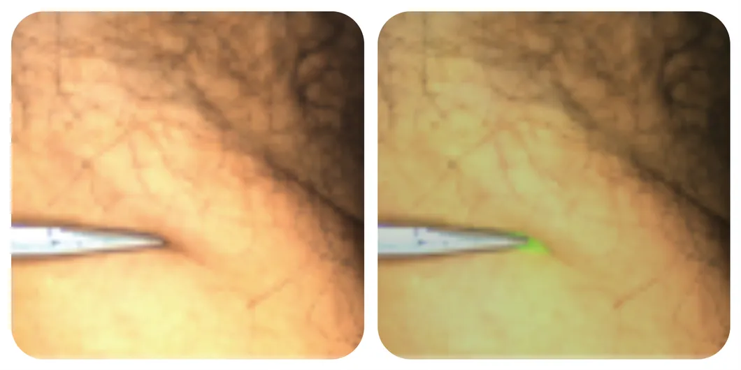
Lower limb lymphatic mapping
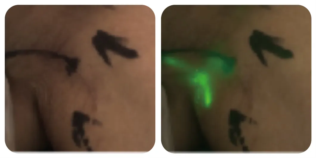
Upper limb lymphatic mapping
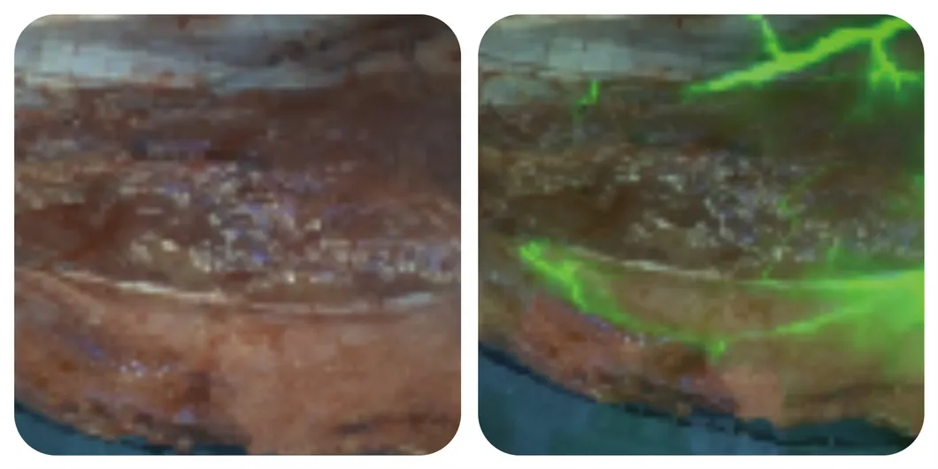
Blood supply evaluation
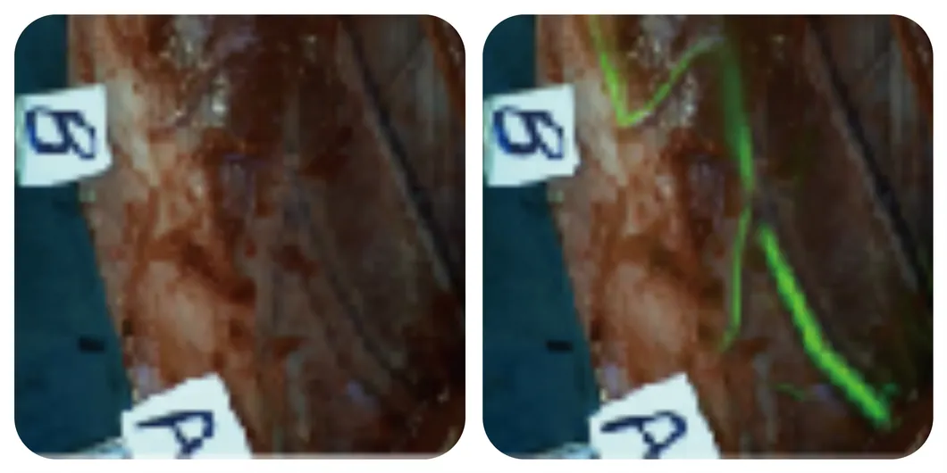
Blood supply evaluation
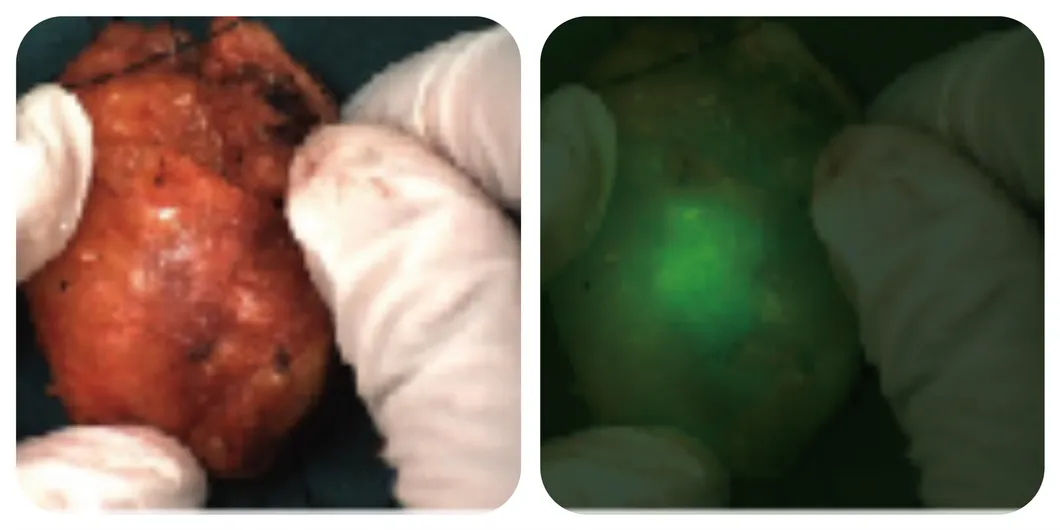
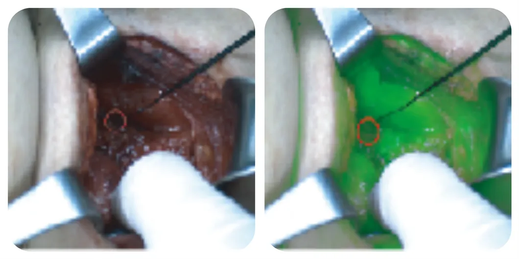
🏥 Precision General Surgery
Used for liver segment staining, biliary tract visualization, and real-time localization of metastases in liver, kidney, and adrenal surgeries.
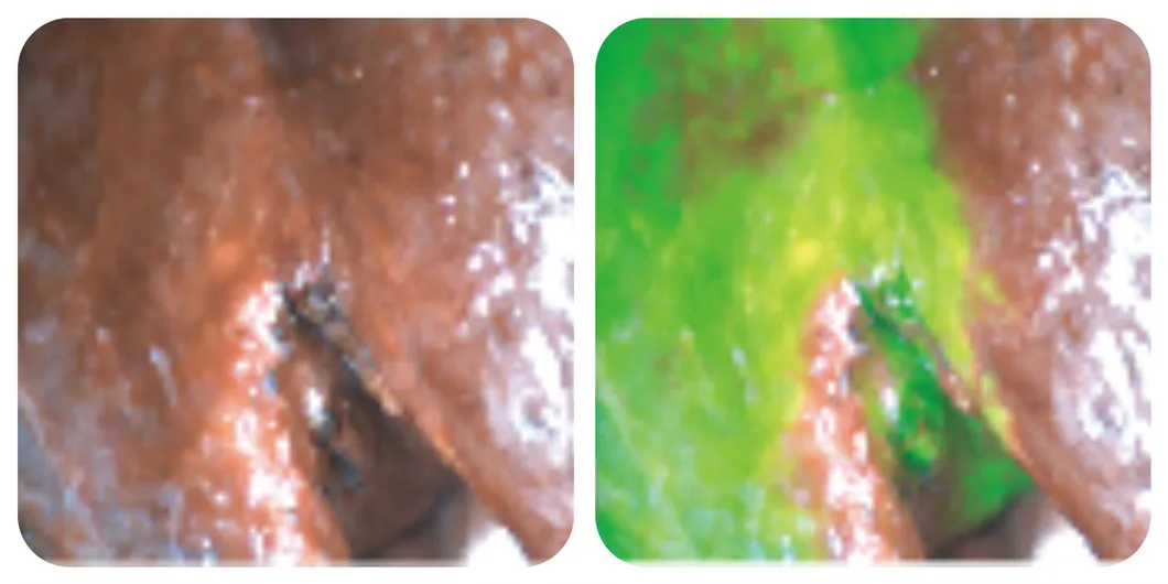
Liver segment staining
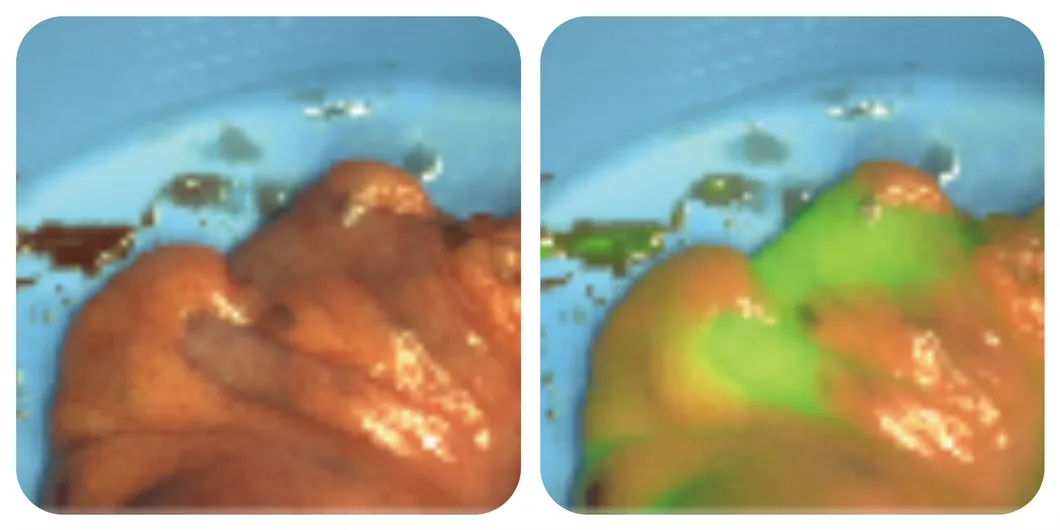
Lymphatic tracing
Tumor identification
Margin determination
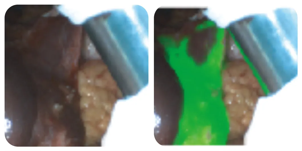
Biliary tract staining
Anastomotic evaluation
Frequently Asked Questions (FAQ)
1. What is the primary use of the Surgical Fluorescence Imaging System?
It is used for real-time surgical guidance, providing high-definition visible light and fluorescence imaging to observe tissues, blood supply, lymph nodes, and anatomical structures during tumor surgeries.
2. Which fluorescence probe is compatible with this system?
The system is designed to work with Indocyanine Green (ICG) as a contrast agent to visualize the circulation of lymphatic systems and blood vessels.
3. What are the key image modes available?
The system offers 7 imaging modes, including White Light, Fluorescence, Fusion, Multimode, Color Grading, Quantification, and Spectroscopy.
4. How sensitive is the system in detecting tumors?
The system features ultra-low fluorescence detection limits (up to 10-12 M/L), enabling the identification of sub-millimeter sized lesions and residual cancer in real-time.
5. Is the system suitable for all types of surgery?
It is specifically optimized for open surgery across multiple departments, including general surgery, oral and maxillofacial, thyroid, breast, and plastic surgery.
6. Does the system provide quantitative data?
Yes, it features a dual quantitative analysis system providing ROI values and independent spectral digitization for objective medical judgments.